- Home
- torn calf muscle
- A novel approach to sonographic examination in a patient with a calf muscle tear: a case report, Journal of Medical Case Reports
A novel approach to sonographic examination in a patient with a calf muscle tear: a case report, Journal of Medical Case Reports
4.9 (456) · $ 25.99 · In stock
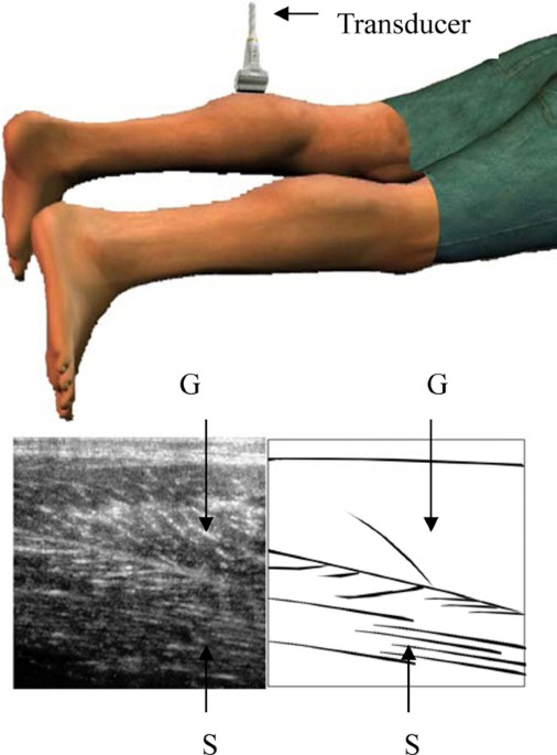
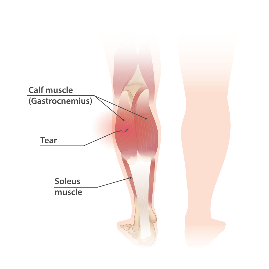
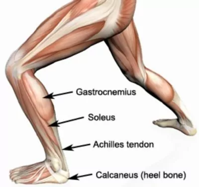
Introduction Rupture of the distal musculotendinous junction of the medial head of the gastrocnemius, also known as "tennis leg", can be readily examined using a soft tissue ultrasound. Loss of muscle fiber continuity and the occurrence of bloody fluid accumulation can be observed using ultrasound with the patient in the prone position; however, some cases may have normal ultrasound findings in this conventional position. We report a case of a middle-aged man with tennis leg. Ultrasound examination had normal findings during the first two attempts. During the third attempt, with the patient's calf muscles examined in an unconventional knee flexed position, sonographic findings resembling tennis leg were detected. Case presentation A 60-year-old man in good health visited our rehabilitation clinic complaining of left calf muscle pain. On suspicion of a ruptured left medial head gastrocnemius muscle, a soft tissue ultrasound examination was performed. An ultrasound examination revealed symmetrical findings of bilateral calf muscles without evidence of muscle rupture. A roentgenogram of the left lower limb did not reveal any bony lesions. An ultrasound examination one week later also revealed negative sonographic findings. However, he still complained of persistent pain in his left calf area. A different ultrasound examination approach was then performed with the patient lying in the supine position with his knee flexed at 90 degrees. The transducer was then placed pointing upwards to examine the muscles and well-defined anechoic fluid collections with areas of hypoechoic surroundings were observed. Conclusion For patients suffering from calf muscle area pain and suspicion of tennis leg, a soft tissue ultrasound is a simple tool to confirm the diagnosis. However, in the case of negative sonographic findings, we recommend trying a different positional approach to examine the calf muscles by ultrasound before the diagnosis of tennis leg can be ruled out.

The Ultrasound Board Review Book is Out! Exclusive POCUS 101 Discount - POCUS 101

If Your MRI Is Normal, Is Your Pain All In Your Head? - Advanced Musculoskeletal Medicine Consultants, Inc.
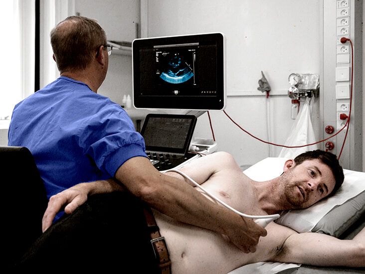
What is an echocardiogram? Uses, procedure, and results

Medical ultrasound - Wikipedia

Chest Pain Differential Diagnosis - The Cardiology Advisor

Sonographic assessment of musculoskeletal causes of calf pain and swelling
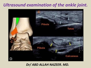
Presentation1.pptx. ultrasound examination of the ankle joint.
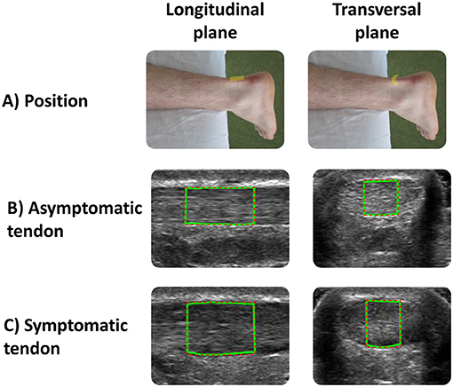
Frontiers To What Extent Do Musculoskeletal Ultrasound Biomarkers Relate to Pain, Flexibility, Strength, and Function in Individuals With Chronic Symptomatic Achilles Tendinopathy?
Cureus, Past and Present of Point-of-Care Ultrasound (PoCUS): A Narrative Review


)